
17100 Wood Acre Trail
Chagrin Falls, Ohio 44023
Phone/Fax: 440-708-2211
Email: BudingerPA@MSN.com
TECHNICAL SERVICE RESPONSE NO.: UT042
Subject: |
Analysis of Black Material on the Roots and Inside Corn Stalks from a Crop Formation
(Mission, B.C., August 2002) |
| Date: |
May 25, 2005 |
| Requested By: |
Nancy Talbott, BLT |
| Reported By: |
P. A. Budinger Analytical Scientist |
Background/Objective:
The background as described by Nancy Talbott follows.
The enclosed corn stalks come from a crop formation in Mission B.C., Canada which
occurred in late August, 2002. You will note that the stalks have a lot of black on them. The
stuff on the leaves I suspect is from fungus. But there is a different quality to the black on
the roots and inside the corn stalks near the bottom which we are wondering about. Could
this be a carbon blackening? Like perhaps one might see in a lightning strike? Or is it just
the fungus?
The objective is to determine the cause of the black color on the ends and roots of the corn stalks.
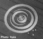
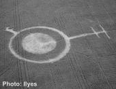
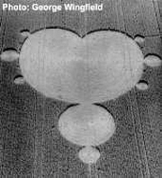
An aerial photograph of the site of the corn formation is below.
·The analysis shows the black interior is corn fungus (ustilago). No carbon, which would be indicative of burning/heat, was detected.
·Two tests for determining the difference between carbon and black corn fungus can be easily done by field workers. The first would be to smell for charred material. The second would involve touching the black area. If the black material is carbon, some will come off and blacken the fingers. Corn fungus would not come off.
Procedure:
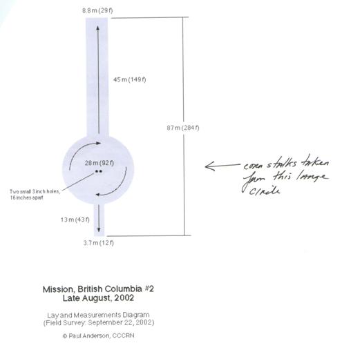
The samples consist of four corn stalks identified as “stalks from crop circle “B” by river”. Following is a diagram of the formation, which shows the two sampling locations of the stalks.
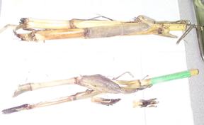 |
The Stalks |
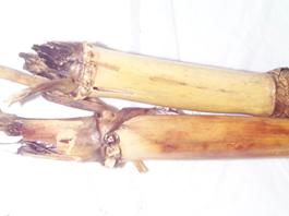 |
Blackened Ends |
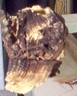 |
End (Close Up) |
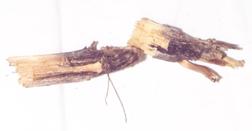 |
Blackened Roots |
On receipt of the sample the black ends and root areas were touched to determine whether any carbon would be transferred to the fingers. None was. The blackened areas were also smelled for the presence of charred material. Again no characteristic odor was detected. This was the first clue that the black material may not be carbon. This was rather surprising to this analyst due to the intense blackness of these areas.
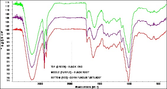
Infrared spectra of the blackened ends and root areas are identical to each other and match a reference of corn fungus (ustilago). No carbon was detected. If carbon were present, the spectra would have displayed significant light scattering in the higher frequency regions (4000 – 1800 cm-1). Following are infrared spectra of the black areas of an end and the root which are compared to a reference of corn fungus. Clearly all spectra are identical.
Infrared Spectra of Corn Stalk Black End and Black Root and a Corn Fungus Reference
And additional note should be made regarding the spectrum of ustilago compared to a ‘clean’ area of the interior of the corn stalk for the scientist familiar with infrared interpretation. The spectra appear to be similar, however, there are significant differences. The ustilago spectrum is clearly typical of fungus. The ustilago fungus spectrum shows: increase in C-H stretch absorption (2000-2800 cm-1); additional ester C=O (1740 cm-1); increased absorption contributed to by an amide C=O (1700-1490 cm-1); increased absorption to a combination of modes (1480-1290 cm-1); loss of resolution in the C-O stretch region (1140-930 cm-1). It is very close to a reference of dried wood ear mushroom which is also a fungus. Following are spectra of the clean corn stalk interior, corn fungus (ustilago), and dried wood ear mushroom illustrating the above.
Infrared Spectra of ‘Clean’ Corn Stalk Interior, Dried Wood Ear Mushroom, and Corn Fungus (Ustilago)

File: UT042
___________________
Phyllis A. Budinger